
If you have a segmental mosaic embryo in storage, the decision is rarely about that embryo in isolation.
You may be trying to understand whether it should be transferred after an available euploid embryo, how it compares with a whole-chromosome mosaic embryo or whether one segmental result may be prioritised over another.
There may also be a wider decision about whether to proceed with transfer or try to create more embryos first.
The comparison begins with understanding what a segmental mosaic result actually means.
What a segmental mosaic embryo result means
PGT-A begins with a biopsy, but the biopsy and the chromosome analysis are two separate stages.
First, an embryologist uses a microscope to remove a small group of cells from the trophectoderm, the outer layer of the blastocyst that forms the basis of the placenta.
The microscope is used to remove the cells. It is not used to examine or count their chromosomes.
The biopsied cells are then sent to a genetics laboratory. Because the sample contains only a tiny amount of DNA, the laboratory first makes many copies of it so there is enough to analyse.
Laboratory equipment then measures how much DNA from each chromosome is present across the whole sample.
When the measurement for part of one chromosome falls between the amount expected when that section is present in all the sampled cells and the amount expected when it is extra or missing from all of them, the laboratory may interpret the result as mosaic.
Mosaic describes the possible mixture in the biopsy:
some cells with the expected amount of DNA from that chromosome section;
some cells with extra or missing DNA from the same section.
Segmental describes how much of the chromosome is involved. The finding affects one section of the chromosome rather than the whole chromosome.
PGT-A does not identify and count the individual cells in each group. The DNA from all the biopsied cells is analysed together, and the possible mixture is inferred from the combined measurement.
This is also why the mosaic percentage on the report is an estimate. It reflects where the combined DNA measurement fell within the laboratory’s reporting scale. It does not mean that the laboratory counted the embryo’s cells and found that an exact percentage carried the segmental finding.
A segmental mosaic result therefore combines two pieces of information: mosaic describes a possible mixture of cells in the biopsy, while segmental means the finding involves part of one chromosome.
How segmental mosaicism differs from whole-chromosome mosaicism
Both results describe a possible mixture within the cells removed during the embryo biopsy. The difference is how much of the chromosome is involved.
A whole-chromosome mosaic result involves the entire chromosome. The combined DNA measurement may suggest that some cells contained the expected two copies of that chromosome while others contained an extra or missing copy.
A segmental mosaic result involves one section of a chromosome. The rest of the chromosome may contain the expected amount of DNA, while part of it appears to be extra or missing in some of the sampled cells.
Published transfer outcomes have generally been more favourable for segmental mosaic embryos than for whole-chromosome mosaic embryos.
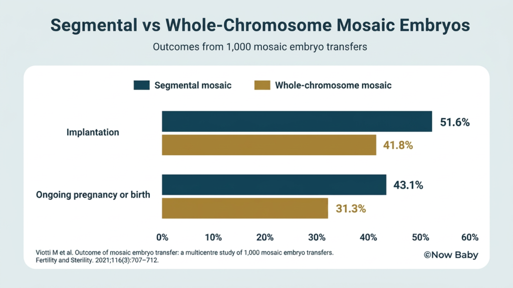
In published studies, segmental mosaic embryos were more likely to implant and lead to an ongoing pregnancy or birth than whole-chromosome mosaic embryos.
This does not create a fixed transfer order. It shows why segmental and whole-chromosome findings should not automatically be placed in the same category.
The next comparison is between different segmental findings, because the amount and location of chromosome material involved also matter.
Why the chromosome segment and biopsy limitations matter
A segmental finding does not carry the same implications in every embryo.
The report may identify the size of the affected section in megabases and show where it begins and ends on the chromosome. Size indicates how much chromosome material is involved. Location identifies which genes and regulatory regions fall within that section.
A larger section may contain more genes, but size alone does not establish its significance. A smaller section may include chromosome material with an important role in development. Two segmental findings of a similar size can therefore carry different clinical considerations because they occur in different locations.
That detail makes the result more specific. The biopsy still places limits on what can be established.
PGT-A analyses a small group of cells removed from the trophectoderm. It can describe the DNA measured in those cells, but it cannot map the chromosome status of every cell in the embryo. The possible mixture may be distributed differently elsewhere in the embryo, and technical variation during the copying and analysis of a very small DNA sample can also contribute to a segmental mosaic result.
The size and location of the segment therefore add important context without turning the biopsy into a diagnosis of the whole embryo. That is why the result needs to be compared with the other embryos available rather than used as a transfer decision on its own.
Why the chromosome segment and biopsy limitations matter
Two embryos can both be reported as segmental mosaic and still raise very different considerations for transfer.
Your report may identify which chromosome is involved, where the affected section begins and ends, and whether part of that chromosome appears to be extra or missing.
The size of the section matters because a larger section usually contains more genetic material. But size cannot establish its importance on its own.
A smaller section may include genes with an important role in early development. A larger section may involve a region where the likely effect is better understood. The chromosome involved and the exact location of the section therefore matter alongside its size.
Genetic counselling can help establish whether the reported section overlaps with a recognised chromosome condition and what is known about the genes within that region.
The report still comes from a small group of cells removed from the trophectoderm, the outer layer of the blastocyst that forms the basis of the placenta. The cells that develop into the baby are not sampled during PGT-A.
The biopsy can show what was measured in those removed cells. It cannot provide a cell-by-cell map of the whole embryo.
The segmental finding may be present elsewhere in the embryo, confined to part of the trophectoderm or influenced by the technical limits of analysing such a small amount of DNA. The report cannot determine which of these explanations applies with certainty.
Transfer priority therefore depends on two parts of the result: what chromosome section was identified and how much weight can reasonably be placed on a biopsy from one small part of the embryo.
Those details can then be compared with the results and grading of the other embryos available to you.
How a segmental mosaic embryo compares with other available embryos
Transfer priority is comparative. It is not a score attached to one embryo in isolation.
If you have an available euploid embryo, it will usually be prioritised before a segmental mosaic embryo. Euploid embryos have produced higher implantation and ongoing pregnancy or birth rates than mosaic embryos in published transfer studies. The result also carries less uncertainty because the DNA measured in the biopsy fell within the expected range.
That does not remove the reproductive potential of the segmental mosaic embryo. It means the euploid embryo generally provides the stronger first transfer option when both are available.
When the remaining embryos are all mosaic, the comparison changes.
Segmental mosaic embryos have generally produced better transfer outcomes than whole-chromosome mosaic embryos. An embryo with one segmental finding may also be assessed differently from an embryo involving several chromosomes or a combination of whole-chromosome and segmental findings.
Embryo grading adds another piece of information. It describes how the blastocyst developed and the appearance of the inner cell mass and trophectoderm under the microscope.
Grading can help distinguish between embryos with otherwise similar PGT-A results. It does not remove or outweigh the chromosome finding. A highly graded segmental mosaic embryo remains segmental mosaic, while a lower-graded euploid embryo remains euploid.
The same segmental mosaic embryo may therefore be transferred next when it is the strongest embryo available, but prioritised after another embryo when a euploid or more favourable mosaic result is in storage.
That comparison becomes more personal once the number and type of embryos remaining are considered together.
Whether creating more embryos is realistic
Once the embryos already in storage have been compared, the remaining decision may be whether to proceed with transfer or try to create more embryos first.
The maternal age relevant to this embryo’s age-related chromosome risk is your age when the egg was collected. Your current age matters because it helps shape what another retrieval may produce now.
AMH, antral follicle count and your previous response to stimulation give your clinic a more individual basis for estimating that prospect. The number of eggs collected, how many fertilised and how many reached the blastocyst stage show what happened across your previous cycle.
Low AMH may mean that fewer follicles are likely to respond to stimulation. It does not determine the chromosome status of an individual embryo, but fewer eggs can mean fewer opportunities to create embryos for testing and transfer.
Your previous cycle may also show where numbers reduced most sharply. A low egg yield, low fertilisation or a substantial drop before the blastocyst stage may mean that creating another transferable embryo could require more than one retrieval.
The decision cannot be based on clinical possibility alone.
Another cycle may be financially out of reach. You may not be willing or able to go through further stimulation, egg collection and waiting. Treatment may already have taken more time, energy or emotional capacity than you can continue to give it.
Future family plans also affect the balance. If you hope for more than one child, creating embryos before transfer may carry more weight. If one pregnancy is now the priority, proceeding with the embryo already in storage may be the more realistic choice.
The decision is whether the prospect of creating more embryos is sufficient to justify what another retrieval would require from you.
Questions to take into genetic counselling before transfer
Genetic counselling should give you a clear explanation of your own embryo report and how it affects the decision in front of you.
Your counselling appointment should cover:
- Which chromosome is involved, and exactly where does the affected section begin and end?
- How much chromosome material is involved?
- Does the result point towards extra or missing chromosome material?
- Is the finding reported as low-range or high-range mosaic, and what cut-offs did this laboratory use?
- How confident is the laboratory that this is a true segmental finding rather than a result influenced by the limits of analysing a very small DNA sample?
- Is the chromosome region linked with a recognised genetic condition, and how much uncertainty remains?
- Is chromosome testing for either genetic parent relevant before making the transfer decision?
- How does this embryo compare with each of the other embryos in storage?
- What factors are likely to influence whether this embryo is transferred?
- What pregnancy testing would be discussed if this embryo leads to a pregnancy, and what could each test establish?
You should leave genetic counselling understanding what was found, what remains uncertain and what factors will shape the transfer decision.
Those answers give you the information needed to decide whether transferring the embryo is the right next step for you.
If you decide to proceed with transfer, the next practical consideration is how to support the biological demands of implantation.
Nutrition support once the transfer decision is made
Not all transfers are successful.
PGT-A helps guide the embryo decision. Once transfer takes place, pregnancy depends on the biological work that follows.
Implantation has 5 distinct phases and each has to complete successfully for pregnancy to continue.
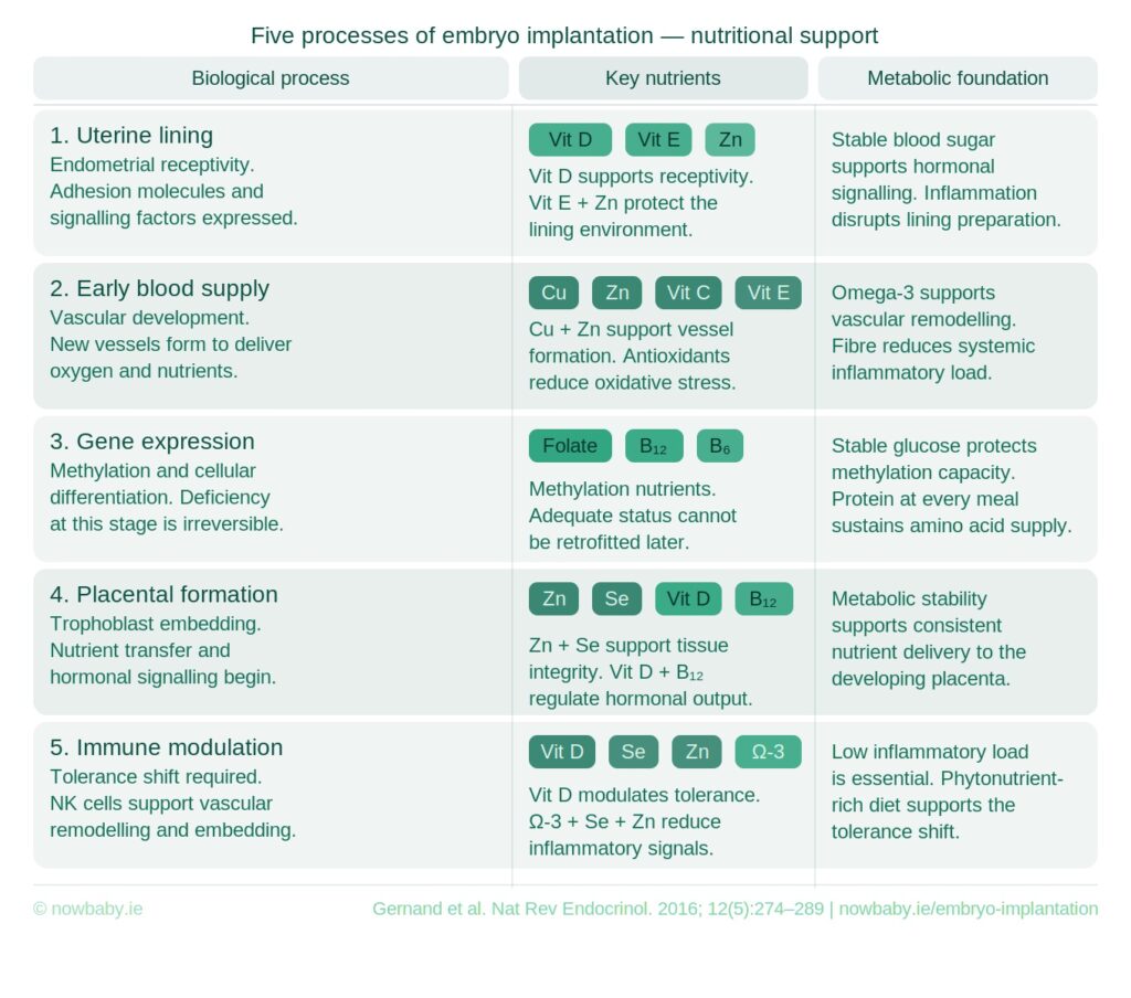
During those stages, cells divide rapidly, genes are regulated, blood vessels begin to develop, the immune system adapts and the earliest placental structures begin to form.
Each of these processes depends on a consistent supply of energy, amino acids, essential fats, vitamins and minerals. Food is how those nutritional raw materials are supplied during the days between transfer and beta.
Your clinic will prepare you meticulously for transfer. You can bring that same level of preparation to meeting the nutritional demands of your embryo at this critical phase.
The Now Baby professionally designed FET Implantation Support Meal Plan takes the guesswork out of this window. It translates the science of the 5-stage implantation process into a structured 14-day food protocol, with every meal designed around the nutritional and metabolic demands between transfer and your beta test.
Get the FET Implantation Support Meal Plan